How do we know whether there is arthritis in a joint from an xray?
Osteoarthritis has a very characteristic appearance on xrays, making it relatively easy to exclude any other types of arthritis.
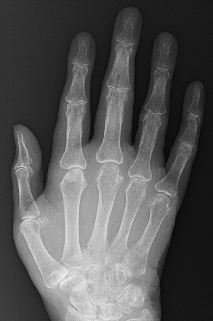
There are 4 features of osteoarthritis on an xray:
– Loss of joint space
– Sub-chondral sclerosis
– Osteophyte formation
– Cyst formation
Hopefully, this page will help you to understand the xrays!
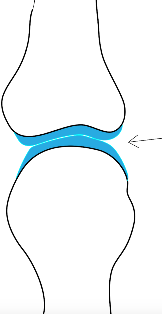
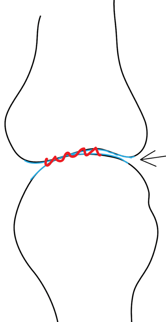
Loss of joint Space
In young patients, there are thick cartilage caps on the ends of the bones within the small joints of the hand. With age, these cartilage caps slowly wear from constant use, despite the body’s regenerative processes. A normal joint, therefore, has much less cartilage at 50 than at 20, and by the time people reach 80+ years, there is often very little cartilage left.
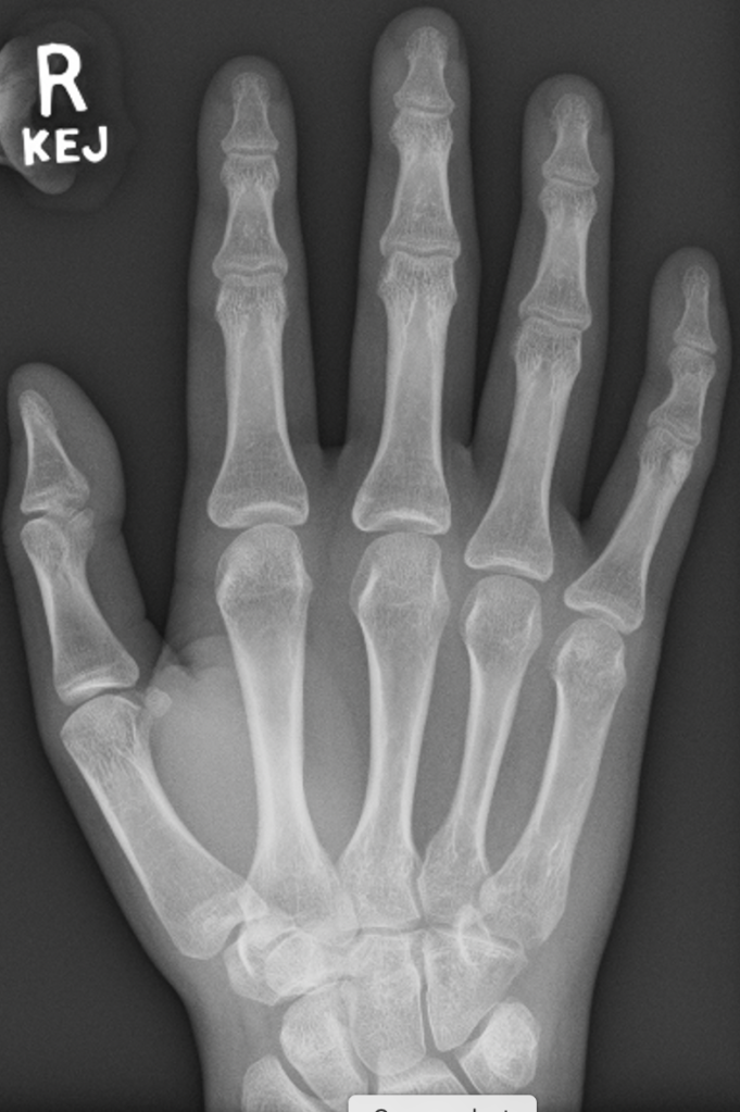
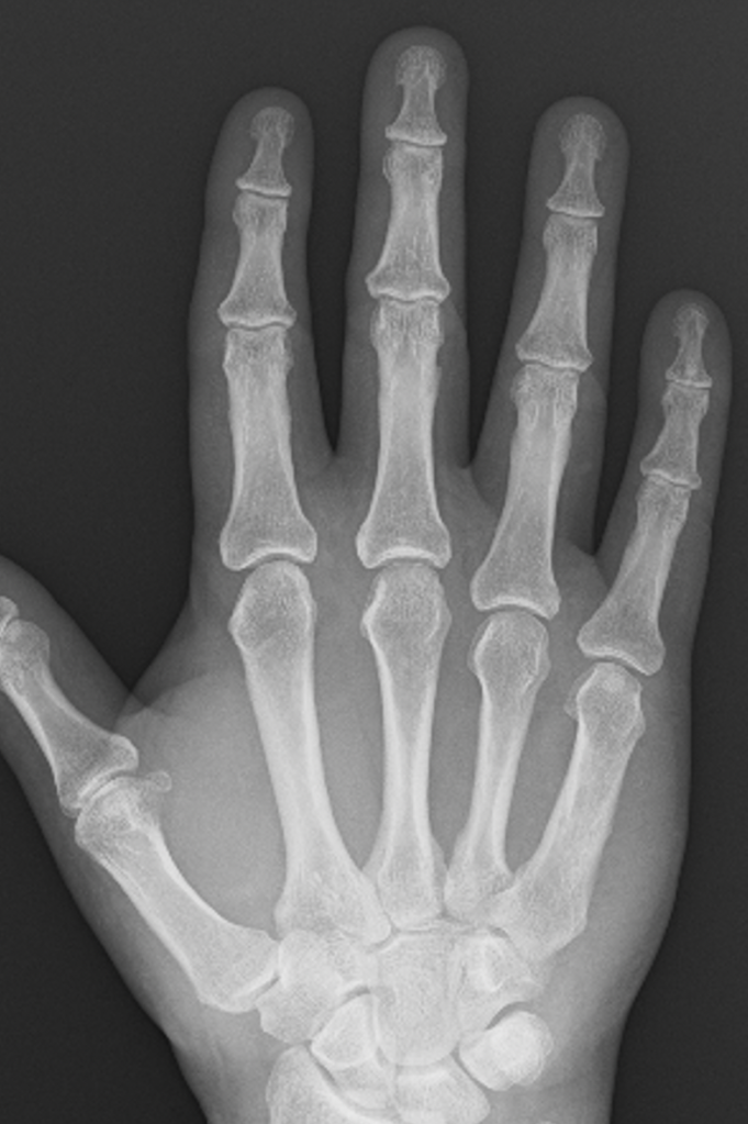
The GAP between the bones on an xray represents the space occupied by the cartilage, and it is very easy to predict how much cartilage is left in a joint simply by looking at the amount of space between the bones – the more space the more cartilage, and generally speaking the more healthy the joint.
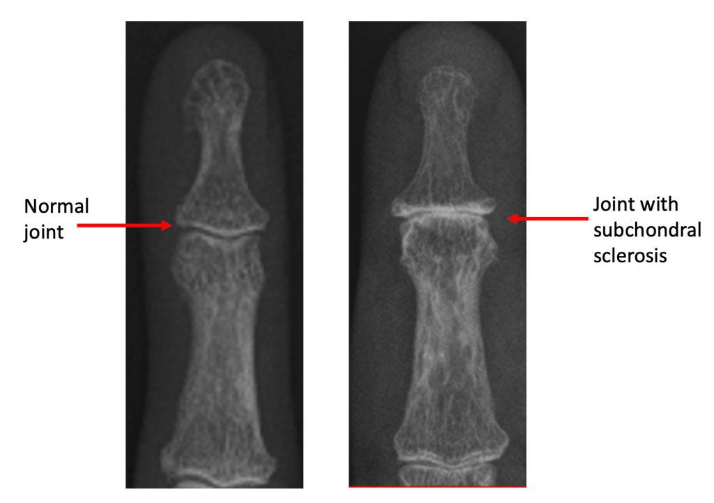
Subchondral Sclerosis
Subchondral simply means ‘under the cartilage‘, and sclerosis is the name given when bone appears thicker or whiter on an xray. When cartilage is lost from the joint, bone articulating with bone triggers a process where the body tries to increase the density of the remaining bone to protect it – on an xray, this gives an appearance of whiteness around the joint, because less xrays can pass through this thickened bone and make it to the xray plate.
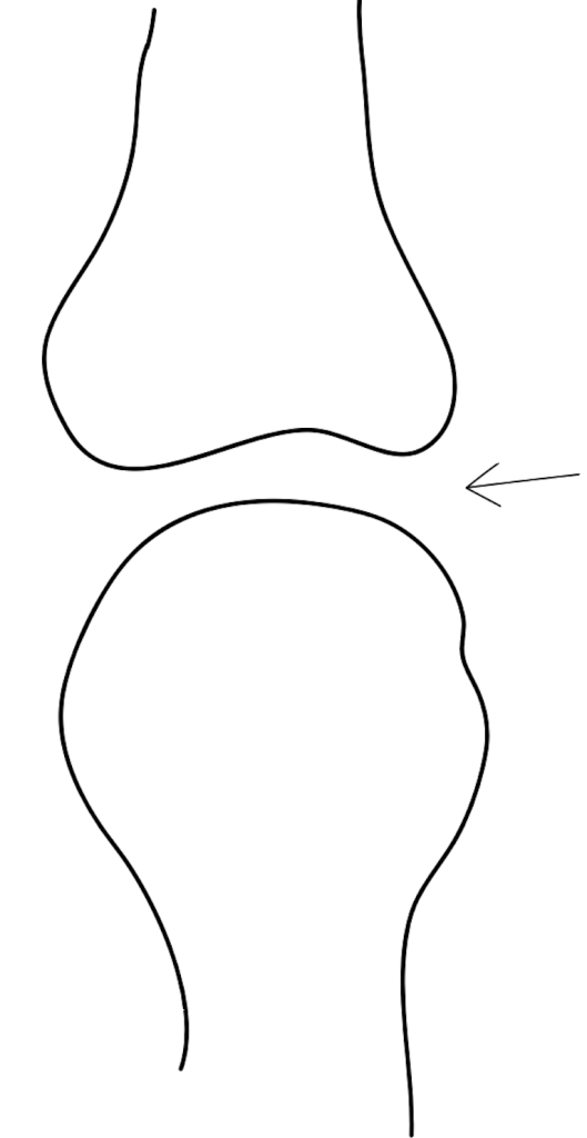
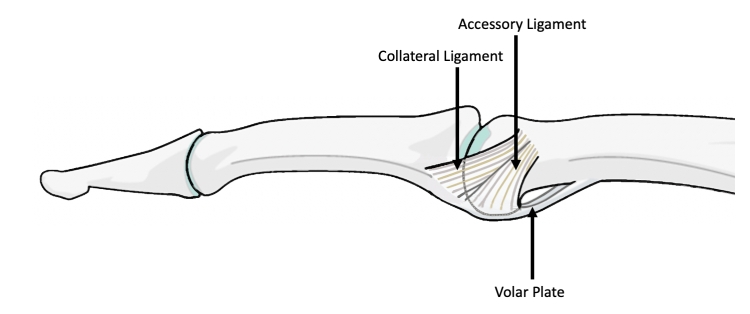
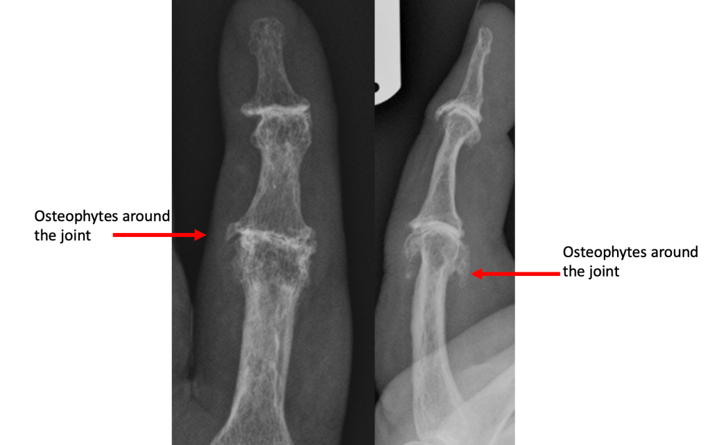
Osteophyte Formation
The stability of a joint depends on the shape and fit of the bone on either side of the joint, and the soft tissues around the joint. In particular, the small joints of the hands have collateral ligaments on either side of the joint, and a tough sheet of fibrous cartilage (volar plate) on the palm side of the joint. These soft tissue structures prevent abnormal movement in the joint.
Where the joint surfaces have worn away, the ligaments and volar plate become slightly too long for the remaining gap, allowing movement of the joint in an abnormal way. The body responds by laying extra bone around the joint in order to make it stable. The extra bone is called osteophyte, and this usually makes the joint stiffen up considerably.
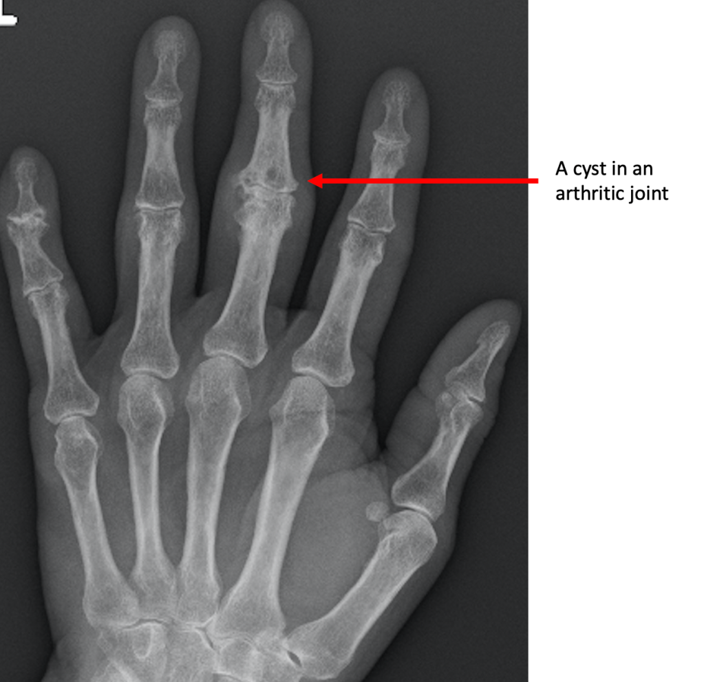
Cyst Formation
Cysts represent areas where the bone has been resorbed or dissolved away by the body. They occur around the arthritic joint, and are thought to occur in response to increased pressure around the arthritic joint. This, in turn, can interfere with the normal blood flow and facilitate cyst formation.
© Aberdeen Hand Service 2021
